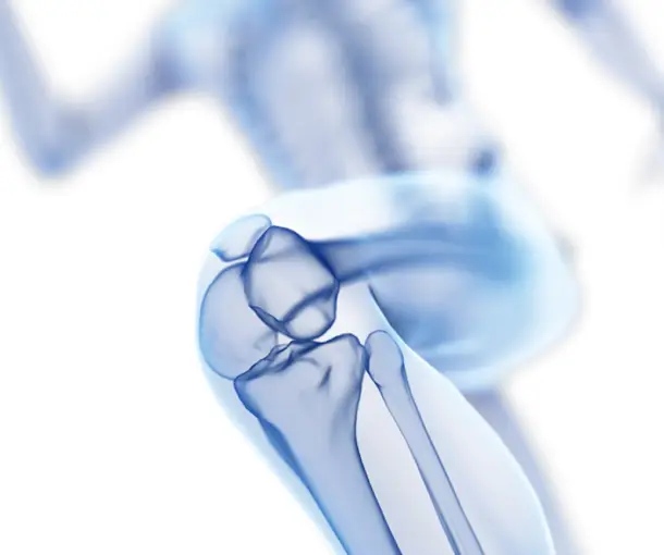
ACL PCL Reconstruction Surgery
What exactly is ACL RECONSTRUCTION?
The anterior cruciate ligament (ACL) reconstruction procedure is used to restore knee stability and strength when the ligament has been damaged.
Why Is ACL Reconstruction Done?
ACL reconstructive surgery is used to repair a damaged ACL and restore knee stability and mobility. While not all instances of a torn ligament need surgery, individuals who are particularly active or who are in constant discomfort may choose to have it done.
ACL repair is often advised if:
– Chronic knee pain.
– Your injury causes your knee to buckle during routine activities, such as walking you are an athlete who wants to remain active
How ACL Reconstruction Is Performed?
The surgical team will be able to provide medicines, anesthetic, or sedatives via the IV. The tendon is then attached with “bone plugs,” or anchor points, to allow it to be grafted into the knee.
During surgery, a small incision at the front of the knee is made to accommodate an arthroscope, which is a thin tube supplied with a fiber optic camera and surgical equipment. During the surgery, your surgeon will be able to view within your knee. The surgeon will first clean the region around your damaged ACL. They will next drill tiny holes in your tibia and femur to accommodate the bone plugs, which will be secured with posts, screws, staples, or washers. Following the connection of the new ligament, the surgeon will assess the range of motion and tension in your knee to verify the graft is stable.
Finally, the opening will be closed, the wound will be bandaged, and your knee will be braced. The duration of the procedure will vary depending on the surgeon’s skill and if other treatments (such as meniscal repair) are undertaken, among other things. Typically, you can go home the day of your surgery.
What are the Risks of ACL Reconstruction?
Because ACL repair is a surgical surgery, there are several hazards involved, including:
– Blood clots and bleeding
– Continued knee pain
– Infection
– Stiffness or weakness in the knees
– Range of motion decrease
– Improper healing if your immune system rejects the transplant
What is Posterior Cruciate Ligament Tear?
The PCL, or Posterior Cruciate Ligament, is one of the ligaments found in the knee joint. It links the thigh or femur bone to the shin bone (tibia). Its primary role is to keep the shin bone from moving rearward during exercise. The PCL tear is a less frequent knee injury caused by a trauma to the front of the knee, particularly when the knee is bent. Injuries might occur as a result of a car accident or while participating in a sport. Partially torn PCLs usually recover on their own.
PCL Treatment Options: Surgical and Non-Surgical
The treatment of posterior cruciate ligament tears is determined on the location of the rupture and the degree of the damage ( grading of the tear ) PCL tears often occur at the following levels:
– Mild (Grade 1) level and
– moderate (Grade 2) level normally recovers on its own and does not need surgery.
– Severe (Grade 3) level and other ligament injuries need surgery to correct the conditions. A serious PCL injury, if not addressed surgically, may result in the beginning of knee joint arthritis at a young age.
– PCL Avulsions needs arthroscopic fixation
Surgical Treatment
If there are several PCL tears or combination injuries, the doctor may propose surgery. On full PCL tears, surgery is generally done only after the swelling has subsided and the knee’s mobility has been recovered, which usually takes a few weeks.
Ligament reconstruction involves replacing the damaged PCL with a new ligament, which is commonly a graft derived from the hamstring, quadriceps, or allograft Achilles tendon. This is accomplished by Arthroscopy, often known as keyhole surgery, in which small incisions around the knee are created and a small camera called an arthroscope is used to see the extent of the damage as well as guide the small specialized equipment required to execute the procedure.
Non-Surgical Therapy
Mild and moderate PCL injuries may heal relatively effectively when treated with non-surgical approaches such as:
RICE: The doctor recommends resting the knee. Gentle compression and elevation of the knee may be required at times for speedier recovery.
Immobilization: The doctor may use a brace to keep the knee from moving. To avoid placing weight on the leg, the patient must walk using crutches.
Physical treatment will be recommended by the doctor after the edema has subsided. The physical therapist will prescribe exercises to restore knee function and strengthen the muscles that support the knee, particularly the quadriceps.
Post – Surgery care
The patient’s recovery following surgery may be affected by the severity of the damage. The combined injuries normally take a long time to heal, although most patients report significant improvement. It may take 3 to 6 months for the patient to fully recover. Physical treatment may be recommended by the doctor to strengthen the knee and restore its mobility.

